
![]()
Research
Overview
People
Profiles
How to Reach Us
Contact Info
Lodish Lab Research Summary
Research in the Lodish laboratory focuses on several important areas at the interface between molecular cell biology and medicine:
We are characterizing many novel genes that are important for terminal stages of erythropoiesis, including gene induction and repression, chromatin condensation, and enucleation. A major focus is identifying genes and extracellular signals that regulate the self- renewal, proliferation, and differentiation of early (BFU-E) erythroid progenitor cells; extracellular signals include activators of the glucocorticoid and PPAR receptors and oxygen. This work has led to the characterization of several molecules, including six that are FDA-approved drugs for other indications that show great promise as therapeutics for bone marrow failure disorders and erythropoietin- resistant anemias. This research involves extensive computational analyses of large datasets of gene expression profiles and chromatin modifications generated from cells at different stages of human and mouse red cell development.
Red cells have a lifespan of 120 days and contain no DNA; any genes introduced into red cell precursors will no longer be present in the enucleated red cells introduced into a recipient. A large DARPA- supported project, in collaboration with Prof. Hidde Ploegh, involves the generation in culture of both murine and human red blood cells that have on their surface monoclonal antibodies that inactivate a variety of toxic substances, or receptors that can bind and remove unwanted materials from the blood. Gene- modified red cells can be targeted to specific sites in the vasculature where they can deliver drugs or reagents or serve as imaging modalities.
C. Long non-coding RNAs (lincRNAs).
We identified many lineage- specific lnc RNAs that are essential for the differentiation and function of erythroid and myeloid cells, as well as others essential for formation of white and/or brown adipose cells. We are identifying their mRNA and protein targets, and studying their roles and mechanisms during cell development and disease.
We are identifying genes and proteins involved in development of insulin resistance and stress responses in adipose cells, including their responses to reactive oxygen species.
What ties all of these projects together is their focus on the basic cell and molecular biology of genes and proteins important for human physiology and disease.
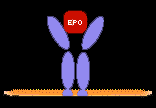
Introduction Erythropoietin (Epo) is the principal regulator of red blood cell production; Epo is produced by the kidney in response to low oxygen pressure in the blood. Epo binds to Epo receptors on the surface of committed erythroid CFU-E progenitors, blocking apoptosis (programmed cell death), their usual fate, and triggering them to undergo a program of 4 - 5 terminal erythroid cell divisions and differentiation. We showed that the first two cell divisions, concomitant with differentiation from CFU-Es to late basophilic erythroblasts, are highly Epo-dependent; differentiation beyond this stage, involving chromatin condensation, ~1-2 terminal cell divisions, and enucleation, is no longer dependent on Epo but is enhanced by adhesion of the cells to a fibronectin matrix. Following condensation of chromatin and subsequent enucleation reticulocytes (immature red cells) are released into the blood. During this terminal differentiation about 400 erythroid- important genes are induced; over many years we have identified many novel transcription factors and cofactors, splicing protein, chromatin modifying proteins, and enzymes in metabolic pathways including heme biosynthesis, that are essential for red cell formation.
An earlier committed erythroid progenitor, termed the burst- forming unit erythroid (BFU-E), can divide and generate additional BFU-Es (that is, undergo self- renewal) or divide and generate later Epo- dependent CFU-E progenitors. Several cytokines and hormones are known to support BFU-E proliferation and formation of CFU-Es, including stem cell factor (SCF, the ligand for the c-kit protein tyrosine receptor) as well as IL-3, IL-6, and IGF-1. However, regulation of BFU-E proliferation, self-renewal, and differentiation during basal and stress conditions is not well understood.
We are focusing much effort in this important area because of the clinical observation that many patients with bone marrow failure disorders such as Diamond-Blackfan anemia are helped by glucocorticoids (GCs) rather than Epo treatment. These patients already have very high Epo levels in the blood, but do not have sufficient Epo-responsive CFU-E cells in the bone marrow to support life.
A culture system that supports normal expansion and terminal differentiation of human hematopoietic stem/progenitor cells
There are many published cell culture systems for expanding human hematopoietic stem and progenitor cells such that they generate hemoglobin- containing nucleated red cell progenitors. But invariably these fail to undergo normal terminal differentiation, condense their nuclei and expel the nucleus from the cell. Jiahai Shi set out to optimize our system of in vitro culture of murine fetal liver Epo- responsive CFU-E progenitors to generate the maximum numbers of enucleated erythroid cells, termed reticulocytes. Careful examination of media constituents led to the discovery that the ferro- transferrin concentration indeed limits terminal erythropoiesis, and supplementation with as much as 500 µg/ml ferrotransferrin greatly enhanced production of reticulocytes. Fetal bovine serum was essential, and ferro transferrin could be partially but not completely replaced by a water- soluble iron chelating agent.
In part based on this work Sherry Lee recently developed a 21-day culture system that supports synchronized erythroid expansion, terminal differentiation, and enucleation of mobilized human peripheral blood CD34+ stem and progenitor cells. She developed a four- stage culture system for these cells that yields a 15,000 to 30,000 fold cell expansion, equal to 14 to 15 cell doublings. At the end of the culture all of the cells were hemoglobinized and thus of the erythroid lineage. Over 45% of the cells had undergone enucleation and each enucleated reticulocyte contained ~30 pg hemoglobin, similar to the amount in each human red blood cell. The hemoglobin composition in these cells was the same as in adult human red cells, and the enucleated reticulocytes averaged 6.7 μm in diameter, similar to that of normal human red blood cells and reticulocytes. Her experiments indicate that timely supply and withdrawal of cytokines required for each developmental stage is important for erythroid differentiation and synchronizing the cell population in culture. She has been conducting further analyses to characterize the proteome and transcriptome of cells at several devlopmental stages.
This culture system enables many studies on terminal differentiation of human erythrocytes. For instance, using lentivirus vectors we have succeeded in expressing one or in some cases two foreign genes in well over 90% of the erythroid cells generated in culture. Similarly, we have used lentiviral vectors expressing shRNAs to knock down expression at will of any erythroid gene. This culture system combined with these manipulations allow unprecedented and focused genetic manipulation of red cells and their major proteins, and allow us to do many experiments on drugs and genes and their effects on red cell formation.
Corticosteroids, hypoxia and stress erythropoiesis
In situations of severe loss of red blood cells mammals respond by a process known as stress erythropoiesis (SE), in which there is increased formation of erythroid progenitors. Glucocorticoids (GCs) are known to be very potent enhancers of SE. This stimulatory effect of GCs on SE is utilized in the therapeutic regimen of Diamond-Blackfan Anemia (DBA), an erythropoietin-resistant congenital red cell aplasia. While an Epo-dependent balance of late red cell precursor survival normally maintains red cell homeostasis, findings by a former postdoc, Johan Flygare, indicate that the physiology of SE involves a stimulation of the earlier BFU-E erythroid progenitors, which when activated are able to rescue red cell production in conditions such as DBA, where erythropoietin has little effect.
Johan showed that glucocorticoids stimulate self-renewal of early Epo-independent progenitor cells (burst-forming units erythroid or BFU-Es), over time increasing production of colony-forming units erythroid (CFU-E) erythroid progenitors from the BFU-E cells, and enhancing the numbers of terminally differentiated red cells. GCs do not affect CFU-E cells or erythroblasts. In mRNA-seq experiments, he found that glucocorticoids induced expression of ~86 genes more than 2- fold in BFU-E cells. Computational analyses indicated that, of all transcription factors, binding sites for hypoxia-induced factor 1 alpha (HIF1α) were most enriched in the promoter regions of these genes, suggesting that activation of HIF1α may enhance or replace the effect of glucocorticoids on BFU-E self-renewal. Indeed, HIF1α activation by the pan-prolyl hydroxylase inhibitor (PHI) DMOG synergized with glucocorticoids and enhanced production of CFU-Es and later erythroblasts over 170-fold. Johan recently established his own research group at the Lund Stem Cell Center in Sweden.
More recently two current postdoctoral fellows, Sherry Lee and Xiaofei Gao, showed that two clinically-tested specific inhibitors of the prolyl hydroxylase that regulates HIF1α activation also synergize with corticosteroids to stimulate both human and mouse BFU-E self renewal and at orders of magnitude lower concentrations than DMOG.
We propose and are testing a physiological model of stress erythropoiesis where increased levels of GCs -systemic stress hormones - and reduced oxygen - local stress - help maintain the earliest erythroid BFU-E progenitors, increase CFU-E output, and at the same time stimulate terminal differentiation, thus promoting both a rapid and long-lasting increase in red blood cell production. Also, PHI-induced stimulation of BFU-E progenitors represents a conceptually new therapeutic window for treating Epo-resistant anemias.
Identifying potential drugs for Epo-resistant anemias that stimulate BFU-E self-renewal and increase production of red blood cells.
Based on our previous study showing that glucocorticoids specifically stimulate self-renewal of BFU-Es and over time increase the production of terminally differentiated red cells, Sherry Lee and Xiaofei Gao tested whether known pharmaceuticals that are either agonists or antagonists of other nuclear receptors affect BFU-E self-renewal and could potentially be used as new therapeutics for anemias that are not treatable by Epo. Using first the mouse fetal liver BFU-E culture system we developed and then our new ex vivo human CD34+ erythroid culture system, they found that two clinically-tested agonists of the peroxisome proliferator-activated receptor alpha (PPARα) synergize with glucocorticoids to promote BFU-E self-renewal and over time greatly increase red cell production. Genome-wide gene expression analyses both in control and corticosteroid- treated mouse BFU-E cells showed that PPARα occupies many chromatin sites that are in close proximity to those occupied by the glucocorticoid receptor (GR), indicating that the GR and PPARα function cooperatively to regulate gene expression.
While PPARα-/- mice show no hematological difference from wild-type mice in both normal and phenylhydrazine (PHZ)-induced stress erythropoiesis, PPARα agonists facilitate recovery of wild-type mice, but not PPARα-/- mice, from PHZ-induced acute hemolytic anemia. Xiaofei and Sherry are currently testing these PPARα agonists on clinically relevant mouse models of Epo- resistant chronic anemias. With the assistance of Russell Elmes, Xiaofei and Sherry have also identified other small molecules including FDA-approved drugs that are effective in promoting erythroid expansion.
A PhD student, Lingbo Zhang, together with Lina Prak, recently completed a high throughput screening of a library of >2000 tested and approved therapeutic compounds for those that can stimulate murine red cell production in culture, either at the BFU-E or CFU-E level, and identified 45 potential "hits." We chose 23 for detailed characterization, mainly on the basis that they were FDA- approved drugs used therapeutically for other indications. Thus far they have shown that four non-steroid drugs and 6 steroid drugs stimulate expansion of murine BFU-E progenitors in culture and stimulate red cell production to the same extent as does the corticosteroid dexamethasone. These drugs also synergize with corticosteroids in stimulating expansion of human erythroid progenitors in culture. Now heading his own laboratory as a Fellow at the Cold Spring Harbor Laboratory, Lingbo is characterizing the actions of each of these drugs in detail, initially on murine cells in culture and increasingly on cultures of differentiating human erythroid progenitors. We are optimistic that this work will identify compounds less toxic than glucocorticoids for treatment of Diamond-Blackfan Anemia and potentially other EPO unresponsive anemias.
The RNA- binding protein Zfp36l2 is required for self-renewal of early erythroid BFU-E progenitors
Lingbo Zhang, together with his technical assistant Lina Prak, identified the RNA- binding protein Zfp36l2 as essential for corticosteroid- induced self-renewal of early burst forming unit-erythroid (BFU-E) progenitors. Lingbo showed that Zfp36l2 is a transcriptional target of the glucocorticoid receptor (GR) in BFU-Es: their Chip-seq experiments showed that the activated glucocorticoid receptor binds to several genomic regions near the transcription start site of Zfp36l2, and luciferase reporter assays demonstrated that these regions are indeed glucocorticoid inducible. These data suggest this gene is a direct transcriptional target of the GR. Zfp36l2 is normally downregulated during erythroid differentiation from the BFU-E stage but its expression was maintained by all tested GR agonists that stimulate BFU-E self-renewal. Knockdown of Zfp36l2 in cultured BFU-E cells completely disrupted glucocorticoid mediated BFU-E self-renewal, but had no effects on cell division rates or cell survival. Lingbo further found that knockdown of Zfp36l2 in transplanted erythroid progenitors prevented expansion of erythroid lineage progenitors normally seen following induction of anemia by phenylhydrazine treatment. Mechanistically, he showed that Zfp36l2 preferentially binds to messenger RNAs that are induced or maintained at high expression levels during terminal erythroid differentiation and negatively regulates their expression levels, including the mRNA for a key transcription factor required for erythroid differentiation. Zfp36l2 therefore functions as part of a molecular switch promoting BFU-E self-renewal and a subsequent increase in the total numbers of CFU-E progenitors and erythroid cells that are generated.
Lingbo Zhang, together with his technical assistant Lina Prak, identified the RNA- binding protein Zfp36l2 as essential for corticosteroid- induced self-renewal of early burst forming unit-erythroid (BFU-E) progenitors. Lingbo and Violeta showed that Zfp36l2 is a transcriptional target of the glucocorticoid receptor (GR) in BFU-Es: their Chip-seq experiments showed that the activated glucocorticoid receptor binds to several genomic regions near the transcription start site of Zfp36l2, and luciferase reporter assays demonstrated that these regions are indeed glucocorticoid inducible. These data suggest this gene as a direct transcriptional target of GR. Zfp36l2 is normally downregulated during erythroid differentiation from the BFU-E stage but its expression was maintained by all tested GR agonists that stimulate BFU-E self-renewal. Knockdown of Zfp36l2 in cultured BFU-E cells completely disrupted glucocorticoid mediated BFU-E self-renewal, but had no effects on cell division rates or cell survival. Lingbo further found that knockdown of Zfp36l2 in transplanted erythroid progenitors prevented expansion of erythroid lineage progenitors normally seen following induction of anaemia by phenylhydrazine treatment. Mechanistically, Zfp36l2 preferentially binds to messenger RNAs that are induced or maintained at high expression levels during terminal erythroid differentiation and negatively regulates their expression levels, including the mRNA for a key transcription factor required for erythroid differentiation. Zfp36l2 therefore functions as part of a molecular switch promoting BFU-E self-renewal and a subsequent increase in the total numbers of CFU-E progenitors and erythroid cells that are generated.
Understanding erythroid self-renewal and fate commitment using single-cell RNA sequencing
All erythroid and megakaryocytic lineage cells are produced by bipotential Megakaryocyte-Erythroid Progenitors (MEPs). Current protocols for isolating these cells have shown that only a small fraction - ~30% of colonies produced by these cells are actually the mixed erythroid-megakaryocyte colonies expected of a bipotential progenitor; most other colonies are entirely composed of megakaryocytes or erythroid cells indicative of unipotential progenitors. A key step, then, is to identify the cell surface markers that can be used to isolate the bipotential cells. Anirudh Natarajan, a new postdoctoral fellow, is using single-cell RNA-seq and computational approaches to address this problem by identifying cell surface proteins likely to be unique to the bipotential progenitors. Following this, he will capture the transcriptomes of these MEP cells as they self-renew or differentiate in culture to erythroid or megakaryocyte progenitors. This will help us understand how lineage restriction from bipotential to unipotent progenitors is established. In addition, he will identify candidate regulators of this process. Experiments perturbing the expression of these genes will identify novel regulators of fate commitment in these bipotential progenitors.
Pathogenic Janus kinase 2 (JAK2) mutant V617F enhances red cell production by triggering CFU-E self-renewal
JAK2-V617F is a mutant activated JAK2 kinase found in most polycythemia vera (PV) patients and those with other myeloproliferative disorders. The mutation enables cytokine-independent activation of JAK2 in cells that express a homodimeric cytokine receptor such as the erythropoietin receptor (EpoR) or related receptors including those for thrombopoietin and G-CSF. JAK2-V617F skews lineage determination of hematopoietic stem and progenitor cells towards the erythroid lineage and increases the number of erythroid progenitors. This leads to overproduction of red cells, consistent with the high percentage of erythroid progenitors from most PV patients that express JAK2-V617F. However, in some PV patients JAK2-V617F is found in only 10-30% of erythroid progenitors, implying that JAK2-V617F might also stimulate terminal erythropoiesis after the erythropoietin (Epo) dependent CFU-E stage.
To confirm this hypothesis, Jiahai Shi showed that expression of JAK2-V617F in murine CFU-Es allows then to divide ~6 rather than the normal ~4 times in the presence of Epo, initially increasing the numbers of CFU-Es and delaying cell cycle exit. Over time the number of red cells formed from each CFU-E is increased ~4 fold; similar to human PV pathology, JAK2-V617F erythroid progenitors eventually differentiate to normal reticulocytes. Microarray analyses comparing JAK2 and JAK2-V617F erythroblasts indicate that JAK2-V617F not only activates EpoR-JAK2 signaling pathways, but also transiently induces non-erythroid-signaling pathways. He showed that purified fetal liver Epo- dependent progenitors express many cytokine receptors additional to the EpoR as well as Stat1 and Stat3 in addition to Stat5, the only STAT normally activated by Epo. JAK2-V617F triggers activation of Stat1 and Stat3, and inhibition of Stat1 by a drug blocks Jak2 V617F mediated erythropoiesis, but does not affect normal erythropoiesis. This abnormal activation of Stat1 and Stat3 leads to transient induction of many genes not normally activated in terminally differentiating erythroid cells and that are characteristic of other hematopoietic lineages. He hypothesizes that these non-erythroid-signaling pathways delay terminal erythroid differentiation and permit extended numbers of cell divisions. These results provide a more complete understanding of PV pathogenesis, in particular in patients in with low numbers of JAK2-V617F expressing erythroid progenitors.
Transcriptional control of gene expression during terminal erythroid differentiation
In the vertebrate, late erythroblasts must undergo terminal differentiation, which involves terminal cell cycle exit and chromatin condensation, to become reticulocytes. In mammals, there is an additional step requiring extrusion of the pycnotic nucleus via an asymmetric cell division. Many aspects of transcriptional regulation of this process remain unknown. Previous RNA sequencing studies on late erythroblasts identified several genes encoding DNA- binding proteins and whose expression is up-regulated during terminal differentiation. Recent results by Xiaofei Gao and Sherry Lee showed that knocking down nuclear coactivator 4 (NCOA4) interrupts the terminal differentiation in both our human CD34 ex vivo differentiation system and cultured mouse fetal liver cells.
Their chromatin immunoprecipitation linked to deep sequencing (ChIP-Seq) results showed that the chromatin occupancy of NCOA4 is enriched at intronic and distal intergenic regions (defined as > 3 kb from transcription start site) in human erythroblasts. Of note, RNA polymerase II proximally occupies more than half of NCOA4-assoiated chromatin sites. De Novo motif discovery of NCOA4-binding chromatin regions also uncovered binding motifs of several transcription factors (TFs) essential for human development. NCOA4 is highly expressed during terminal erythroid differentiation and knocking down NCOA4 significantly impaired human erythroid terminal differentiation. Ncoa4 KO mice are also severely anemic suggesting a conserved role of NCOA4 in vertebrate erythropoiesis.
Thyroxin regulates several stages in erythropoiesis
While effects of the thyroid hormone thyroxin (TH) on erythropoiesis have been known for more than a century, the molecular mechanisms by which TH affects erythropoiesis remained elusive. Xiaofei Gao found that thyroid hormone is necessary for terminal erythroid differentiation; removal of lipophilic molecules from serum used in our cultures blocked terminal red cell differentiation and enucleation, and addition just of thyroxin resulted in normal differentiation. Xiaofei next showed that the thyroid hormone receptor β (TRβ) directly binds to regulatory segments of many erythroid genes and regulates their expression. Further, TRβ interacts with NCOA4 and genome wide analysis suggests that T4 recruits NCOA4 to the RNA Polymerase II (Pol II) complex, which together occupy genomic regulatory sites encoding abundant transcripts during terminal erythroid differentiation. Collectively, these results not only reveal a novel role of NCOA4 during erythroid terminal differentiation but also uncover the molecular mechanism of thyroid hormone function on red blood cell formation and are useful for developing strategies to treat anemias.
Transcriptional divergence and conservation of human and mouse erythropoiesis
Mouse models have been used extensively for decades and have been instrumental in improving our understanding of mammalian erythropoiesis. Nevertheless, there are several examples of variation between human and mouse erythropoiesis. In collaboration with Vijay G. Sankaran, a recent postdoc and currently an Assistant Professor at Boston Children's Hospital, Nova Pishesha performed a comparative global gene expression study using publicly available data from morphologically identical stage-matched sorted populations of human and mouse erythroid precursors from early to late erythroblasts. Surprisingly, they found that, at a global level, there is a significant extent of divergence between the species, both at comparable stages and in the transitions between stages. This was especially the case for the 500 most highly expressed genes during development, save for some major transcriptional regulators of erythropoiesis and major erythroid-important proteins. This suggests that the response of multiple developmentally regulated genes to key erythroid transcriptional regulators represents an important modification that has occurred in the course of erythroid evolution. They further developed this compendium of data as a systematic framework that is very practical and useful to understand and study conservation and divergence between human and mouse erythropoiesis as well as to help translate findings from mouse models to potential therapies for human disease.
The pre-mRNA splicing factor Muscleblind-like 1 (Mbnl1) regulates pre-mRNA alternative splicing during terminal erythropoiesis
The scope and role of regulated exon use in pre-mRNAs during erythroid development is poorly understood. Using their mRNA- seq data sets from erythroid progenitors and mature Ter-119+ erythroblasts, Jiahai Shi and Bill Wong, working with Albert W. Cheng in Prof. Chris Burge's lab and UROPs Katherine Luo, Paula Trepman, and Heejo Choi, identified hundreds of differentiation-associated isoform changes during terminal erythropoiesis. During differentiation there were major changes in eight classes of alternative isoform expression events involving alternative splicing, alternative 3' end cleavage and polyadenylation, and/or alternative promoter usage; these included skipped exons, mutually exclusive exons, alternative 5' and 3' splice sites, alternative first exons, alternative last exons, tandem 3' untranslated regions, and retained introns. They focused on alternative exon use during differentiation; many of these changes in usage coincided with induction of ~400 erythroid-important genes as well as repression of about 6000 early- stage genes, suggesting that both large-scale transcriptional and post-transcriptional programs are critical to ensure proper erythroid differentiation.
Segments in pre mRNAs surrounding regulated exons were enriched in motifs corresponding to the splicing factor, muscleblind-like1 (Mbnl1). Knockdown of Mbnl1 in cultured murine fetal liver erythroid progenitors resulted in a strong block in erythroid differentiation and disrupted the developmentally regulated exon skipping of several pre mRNAs, including Ndel1 mRNA, which they showed is a direct target of Mbnl1. Knockdown of Ndel1 also impaired erythroid terminal proliferation and was partially rescued by the Ndel1 inclusion form, alternative splicing isoform upregulated during terminal erythropoiesis. These findings reveal an unanticipated scope of the alternative splicing program and the importance of Mbnl1 and Ndel1 during erythroid differentiation.
Translational control of red cell development
Wenqian Hu is investigating how mRNA-binding proteins regulate erythroid terminal differentiation. He characterized one such protein, Cpeb4, that is required for terminal erythropoiesis. Specifically, he found that Cpeb4 is dramatically induced during erythroid terminal differentiation by the two erythroid-important transcription factors, Gata1 and Tal1. Knocking down this protein inhibits this cell differentiation process. Interestingly, Cpeb4 interacts with eIF3, a general translation initiation factor, to repress the translation of a large set of mRNAs in terminal differentiating erythroblasts, including its own mRNA. Thus, transcriptional induction synchronizes with translational repression to maintain Cpeb4 protein within a specific range during terminal erythropoiesis; this precise control of gene expression is required for normal cell differentiation.
Genes important for red cell formation identified by genetic analyses of human erythropoiesis
Vijay Sankaran along with laboratory colleagues Leif Ludwig, Jenn Eng and Hyunjii Cho have been dissecting the genetic architecture of human erythropoiesis, along with colleagues at the Broad Institute. This work is being performed using a combination of complex trait genetics, Mendelian genetics, and analysis of rare human syndromes.
Regulation of fetal globin expression
Elevated levels of fetal hemoglobin can ameliorate the major disorders of beta-hemoglobin diseases, sickle cell disease and beta-thalassemia in particular. They followed up on a several decades old observation that patients with trisomy 13 have elevated levels of fetal hemoglobin and used mapping of partial trisomy cases to show that elevated levels of microRNAs 15a and 16-1 appear to mediate this phenotype. A direct target of these microRNAs, MYB, plays an important role in silencing the fetal and embryonic hemoglobin genes. Thus they have demonstrated how the developmental regulation of a clinically important human trait can be better understood through the genetic and functional study of aneuploidy syndromes, and suggest that miR-15a, 16-1, and MYB may be important therapeutic targets to increase HbF levels in patients with sickle cell disease and β-thalassemia. Following up on this work, this group is defining the physiological function of these microRNAs and their targets using a variety of approaches in primary mouse and human erythroid progenitor cells. Ongoing work is aimed at understanding the mechanistic basis for alterations in hemoglobin expression in the context of other rare human syndromes and clinical conditions.
Cyclins that regulate proliferation of red cell progenitors and red cell size
Using complex trait genetics, this group has been defining new regulators of human erythropoiesis. By using readily measured erythrocyte traits and following up on the results of genome-wide association studies (GWAS), new mechanisms underlying the regulation of erythropoiesis are being defined. Using such approaches, they have recently defined a role for the pleiotropic cell cycle regulator, cyclin D3, in regulating the number of divisions that occur during terminal erythropoiesis, thereby controlling erythrocyte size and number. Specifically, this GWAS variant affects an erythroid-specific enhancer of CCND3. A Ccnd3 knockout mouse phenocopies these erythroid phenotypes, with a dramatic increase in erythrocyte size and a concomitant decrease in erythrocyte number. By examining cultures of differentiating human and mouse primary erythroid progenitor cells, they demonstrated that the CCND3 gene product, cyclin D3, regulates the number of cell divisions that erythroid precursors undergo during terminal differentiation before enucleation, thereby controlling erythrocyte size and number. Similar findings have identified cyclin A2 as a novel regulator of red blood cell size. Ongoing studies are aimed at broadening these approaches to other loci across the genome.
Diamond-Blackfan anemia
Finally, to gain further insight into important regulators of erythropoiesis, this group has been using Mendelian genetic approaches to identify genes involved in erythropoiesis that are mutated in rare human diseases. With collaborators at a number of institutions, new candidate genes mediating these diseases have been defined and functional work is being performed to understand the nature and mechanism of action of these genes. For example, with close collaborator Dr. Hanna Gazda, this group has recently defined the first non-ribosomal protein gene involved in Diamond-Blackfan anemia, GATA1. Diamond-Blackfan anemia is more commonly caused by heterozygous deletions or loss of function mutations in one of 12 ribosomal protein genes, but it was not clear how mutations in ubiquitously expressed ribosomal protein genes could result in an erythroid-specific defect. Further work showed that translation of GATA1 mRNA is impaired in the setting of ribosomal haploinsufficiency. Since GATA1 is essential for erythropoiesis, this provided compelling evidence about the specificity of the defect observed in patients with Diamond-Blackfan anemia with ribosomal gene mutations. When studying the transcriptional signature in stage-matched sorted cells obtained directly from patients with RPS19 mutations, GATA1 target genes were significantly downregulated and GATA1 overexpression can partially rescue defects in primary cells from patients with Diamond-Blackfan anemia. These observations illuminate the central role of GATA1 in the pathogenesis of Diamond-Blackfan anemia. Ongoing studies are defining the mechanisms by which this and other mutations affect human erythropoiesis.
Carrying on a 45-year-old tradition of close interactions and scientific exchanges between the Division of Hematology/Oncology at Boston Children's Hospital and the Lodish laboratory (that began when Dr. David Nathan was a sabbatical visitor with Harvey in 1970), Vijay is continuing this work in his laboratory at Boston Children's Hospital as an Assistant Professor of Pediatrics at Harvard Medical School.
Congenital Dyserythropoietic Anemia (CDA)
Insight into the rare disease Congenital Dyserythropoietic Anemia (CDA) is further elucidating our understandings of erythropoiesis. Unlike Diamond-Blackfan Anemia where there is a lack of erythroid progenitors, patients with CDA have bone marrow hyperplasia, but a derailment in later erythropoiesis causes a decreased output of mature red blood cells. In recent years the genetic cause of the most common CDA subtype, CDAII, was discovered to be caused by a mutation in SEC23b, implicating it in a vesicle transport defect. Recently two siblings with an unusual form of CDA were mapped by Vijay Sankaran to a different genetic locus. In collaboration with Vijay Sankaran's lab at Boston Children's Hospital, Jenn Eng is performing experiments to follow-up on exome sequencing on the effected siblings and their unaffected parents in order to gain a better understanding of what is causing their CDA and what clinical implications this may have for understanding late erythropoiesis.
Modeling disorders of erythropoiesis in primary human and mouse erythroid cells
In ongoing work by post-doc Hojun Li, in collaboration with Drs. Dan Bauer and Matt Canver from Stuart Orkin's laboratory at Boston Children's Hospital, he is attempting to model human diseases of erythropoiesis in our human erythroid culture system. Hojun is utilizing the CRISPR/Cas9 nuclease system to target various genes known to be mutated in human anemias, and is working with Sherry Lee's CD45 cell culture system to determine the stages of erythropoiesis are affected by these mutations. Hojun has also adapted the CRISPR/Cas9 system to the mouse fetal liver erythroid progenitors and demonstrated efficient gene disruption resulting in loss of protein expression. He and Jiahai Shi are planning to use this system for high-throughput screening of novel regulators of erythropoiesis.
Chromatin condensation and enucleation
Mammalian erythroid cells undergo enucleation during a late stage of differentiation, a process that does not occur in other vertebrates. This process has critical physiological and evolutional significance for the morphogenesis and hemoglobin enrichment of mature mammalian red blood cells. Although enucleation has been known for decades, the mechanisms that regulate this process remain obscure. Peng Ji began studying enucleation in our lab, and identified key roles for Rac GTPase and fir the formin (actin nucleating protein) mDia2 in the final step of erythroblast enucleation - the formation of the contractile actin ring on the plasma membrane of late-stage erythroblasts at the boundary between the cytoplasm and nucleus of enucleating cells.
In collaboration with Tzutzuy Ramirez and Junxia Wang, fellows with Maki Murata Hori of the Temasek Life Sciences Laboratory, Singapore, Peng investigated the roles of many cytoskeletal and other proteins in nuclear migration and enucleation of these cells, in part using video microscopy of cells expressing fluorescent- tagged proteins. Initial results show that, unlike conventional cytokinesis, the nucleus is squeezed out by formation of a bleb-like protrusion from a limited area of the erythroblast cell cortex; the bleb increases in size by dynamic contractions of asymmetrically distributed actomyosin. Importantly, they showed that enucleation requires establishment of cell polarization that is regulated by microtubule-dependent local activation of phosphoinositide 3-kinase (PI(3)K), displacing the nucleus to one side of the cell, and restricting actin to the other side. Peng is continuing to work on these and related projects in his new position as Assistant Professor of Pathology at the Northwestern University Medical School.
Histones to the cytosol: Exportin 7 is essential for erythroid nuclear condensation and enucleation.
Together with her undergraduate student, Austin Gromatzky, Shilpa Hattangadi has begun to uncover the function of an unusual regulator of erythroid chromatin condensation and enucleation, the nuclear export protein, Xpo7. Xpo7 is highly erythroid specific and induced markedly during terminal differentiation; its expression is regulated by master erythroid transcriptional regulators. It is unusual for a nuclear export protein in that it does not require a specific nuclear export signal, as do all other exportins. Interestingly, except for Xpo7 transcripts of all other nuclear exportins are repressed during terminal erythropoiesis. Shilpa discovered that erythroblast nuclei from Xpo7- knockdown cells were less condensed and larger than control nuclei, as judged by confocal immunofluorescence microscopy. Enucleation was blocked, and Xpo7- knockdown nuclei retained almost all nuclear proteins while normal extruded nuclei had very little protein, as judged both by silver stained gels and mass spectrometry. This suggested that Xpo7 is a nonspecific nuclear export protein that removes all nuclear proteins from the erythroid nucleus in order to allow chromatin to condense. Strikingly, DNA binding proteins such as histones H2A and H3 accumulated in the cytoplasm of normal late erythroblasts prior to and during enucleation, but not in Xpo7-knockdown cells. Thus chromatin condensation during erythroid development involves removal of histones from the nucleus facilitated by Xpo7. Along with Austin, she is using immunoprecipitation and yeast-2-hybrid methods to uncover the erythroid-specific cargos of Xpo7. She is continuing this work as Assistant Professor in the Departments of Pediatrics and Pathology at Yale University School of Medicine.
Dynamics of the nuclear lamina during terminal erythropoiesis
Chromatin condensation during terminal differentiation is accompanied by proportional shrinkage in the size of the nucleus. The nuclear lamina is composed of the fibrous proteins nuclear lamin A/C and nuclear lamin B, and forms a dense fibrillar network on the inside the nuclear envelope that provides structural support to the nucleus. Jiahai Shi and undergraduate Heejo Choi showed that the expression of the three lamins initially increased and then decreased markedly during terminal erythroid development, and that the dynamic expressions of Lamin A/C and Lamin B controls the thickness of the nuclear lamina. Unlike the gradual decrease in nuclear size, the nuclear lamina increases dramatically in the early stages of terminal erythropoiesis, followed by a sudden decrease at the end, as revealed both by western blotting and immunofluorescence confocal microscopy. These results suggest that enhanced expression of lamin A/C may be important for erythroid terminal differentiation. Jiahai and Heejo are continuing to explore the functional role of the up-regulation of nuclear lamina in terminal erythropoiesis.
Membrane protein sorting during enucleation
Certain membrane proteins are selectively retained on the red blood cell membrane and others in the erythroblast are lost; this selection process mainly occurs during enucleation when nuclei are expelled, surrounded by a segment of the erythroblast plasma membrane, and separated from the remaining reticulocyte. As examples, Glycophorin A, protein 4.1 and Kell protein are retained on the surface of the reticulocytes whereas erythroblast macrophage protein and transferrin receptor (Tfr) are extruded with the nuclei. Proper sorting of membrane proteins at the end of erythroid differentiation is essential for generating normal red blood cells but the regulation and mechanisms of protein sorting before and during enucleation are poorly understood. Nai-Jia Huang is beginning to investigate the mechanisms of protein selection during enucleation. Understanding these mechanisms might provide a new way to express proteins on red blood membranes without modifying endogenous proteins.
| B. Red blood cells as vehicles for the introduction of novel therapeutics, immunomodulatory agents, and diagnostic imaging probes into the human body. |
Red blood cells possess many unique characteristics that make them attractive candidates for in vivo delivery of natural and synthetic payloads. They have a long circulatory half-life (~120 days in humans and ~50 days in mice), and old or damaged RBCs are removed and degraded by cells of the reticuloendothelial system. They are biocompatible and have a large surface area of ~ 140 µm2 with a favorable surface to volume ratio. Importantly, they contain no DNA; any genes introduced into red cell precursors will no longer be present in the enucleated red cells introduced into a recipient.
A large DARPA- supported project, in collaboration with Prof. Hidde Ploegh, involves the generation in culture of both murine and human red blood cells that have on their surface monoclonal antibodies that inactivate a variety of toxic substances, or receptors that can bind and remove unwanted materials from the blood. Gene- modified red cells can be targeted to specific sites in the vasculature where they can deliver drugs or reagents or serve as imaging modalities.
Engineered erythrocytes generated by sortase-mediated modification ("sortagging") of erythroid membrane proteins.
In collaboration with Dr. Lenka Kundrat and other members of Prof. Hidde Ploegh's laboratory at the Whitehead Institute, Jiahai Shi, Sherry Lee, Nova Pishesha, and Nai-Jia Huang are generating specific types of genetically engineered human and mouse erythrocytes (eRBCs) in culture, and are using the sortase technique to covalently link proteins and small molecules to these eRBCs which then can be used for multiple types of applications. Sortases are transpeptidases derived from bacteria - they are involved in cell wall biogenesis - that normally link together two proteins that contain at their ends particular short amino acid sequences. While the sortases from different bacteria recognize different sequences, the most commonly used sortase cleaves between the T and G residues in the flexible motif LPXTG, generating a thioacyl enzyme intermediate at the C-terminus of the threonine residue. This is resolved by nucleophilic attack in a reaction that involves an N- terminal glycine-initiated peptide or probe, (G)n-Y, where n =1 - 5. Therefore, the sortase-based approach (sortagging) covalently links the peptide-based motifs, X-LPXTG and (G)n-Y, resulting in a new structure: X-LPXT(G)n-Y, where X or Y could be any protein, peptide, high molecular weight polymer, or small molecule. Sortase accommodates a wide range of natural and synthetic payloads that allow modification of RBCs with substituents that cannot be encoded genetically.
As proof of concept, Jiahai, Sherry, Nova, and Lenka expressed the sortaggable versions of two erythroid membrane proteins - Glycophorin A, a Type I transmembrane protein, and the Type II transmembrane protein Kell, on the surface of murine red cells made in culture. The expression of sortaggable Glycophorin A was sustained as erythroid differentiation proceeded; the cells underwent terminal erythroid differentiation and could readily be sortagged with a biotin label at very high efficiency. They also generated, using Cas9 technology, a line of mice in which all Kell proteins contained the sortaggable LPXTG motif at its C- terminus; these mature red cells could be sortagged at high efficiency and, importantly, the modified red cells remained in the bloodstream for up to 28 days. A single domain antibody attached enzymatically to RBCs enabled them to bind specifically to target cells that express the antibody target. They extended these experiments to human red cells and demonstrated efficient sortase-mediated labeling of in vitro differentiated human reticulocytes.
Nova Pishesha is currently exploring the possibility of applying this method to modulate the immune response. There are many other ways to use these sortase- and genetically modified erythrocytes produced in culture; initially we will focus on linking fibrinolytic proteins to red cells that could be used therapeutically. The engineered red cells with fibrinolytic protein, like plasminogen activators (PA), should have a long half-life in the circulation. These cells will aggregate around a nascent blood clot, as would normal erythrocytes, and increase the local PA concentration to dissolve the blood clot. This therapeutic should be very useful in preventing thrombosis in patients having a high risk for life-threatening thrombosis like coronary thrombosis. Importantly, there are many other potential uses that we will explore as time permits, including novel therapeutics, stabilizing otherwise unstable red cells, novel immune modulators and vaccines, and novel imaging modalities.
Engineered red blood cells that bind toxic proteins
Nai-Jia Huang and Nova Pishesha are testing whether a chimeric protein, fusing a single domain antibody with either glycophorin A or Kell can be used for expressing these molecules on the red blood cell surface. Single domain antibodies (VHH) are the antigen- binding domains of the unique functional heavy- chain- only camelid antibodies. These VHHs have equivalent binding activities to their cognate antigens compared to conventional IgGs and these VHHs are more stable and easier to make. VHHs that can target botulinum neurotoxin have been discovered and studied intensively by our collaborator Dr. Charles B. Shoemaker of Tufts University Veterinary School; these can neutralize botulinum neurotoxins in cell culture and in vivo. Chimeric genes fusing these VHHs sequences with glycophorin A or Kell cDNA are being assembled and these chimeric proteins will be expressed on mouse and human erythroid cells. Surface expression of these chimeric proteins and abilities neutralize neurotoxins will be examined in collaboration with Dr. Shoemaker.
Long non-coding RNAs (lncRNAs) are transcripts longer than 200nt that do not function through encoded protein products. Many are capped, polyadenylated, and often spliced, and transcribed by RNA Polymerase 2 (Pol2). lncRNAs constitute a significant fraction of the mammalian transcriptome. Compared to mRNAs, lncRNAs tend to be shorter and less well conserved at the primary sequence level. Expression of lncRNAs is often restricted to specific tissues and developmental stages, suggesting that many may regulate cell fate specification .A few dozen intergenic lncRNAs (lincRNAs) have been functionally characterized in mammals, and they have been associated with important developmental processes such as apoptosis, proliferation, and lineage commitment. However, the biological functions of most of these genes and their potential roles in disease still remain uncharacterized.
An erythroid-specific long non-coding RNA prevents apoptosis of erythroid progenitors and promotes terminal proliferation.
Although the regulation of erythropoiesis by transcription factors and microRNAs is becoming well understood, the modulation of red blood cell development by lncRNAs is still unknown. LncRNAs can regulate gene expression via multiple mechanisms and many lncRNAs are differentially expressed in many developmental and pathological processes, suggesting that they play important biological roles.
Wenqian Hu identified one erythroid-specific lncRNA, LincRNA-EPS, with potent anti-apoptotic activity. Expression of LincRNA-EPS is largely confined to terminally differentiating fetal erythroid cells and its expression is induced in CFU-E progenitors by Epo. Inhibition of this lncRNA blocks erythroid differentiation and promotes apoptosis. Ectopic expression of this lncRNA in CFU-E progenitors prevents erythroid progenitor cells from the apoptosis that is normally induced by erythropoietin deprivation. This lncRNA represses expression of several proapoptotic genes including the one encoding Pycard, an activator of caspases, explaining in part the inhibition of programmed cell death. These findings reveal a novel layer of regulation of cell differentiation and apoptosis by a lncRNA. Currently Wenqian, together with Juan R. Alvarez-Dominguez are identifying and cloning the human LincRNA-EPS ortholog and characterizing its putative antiapoptotic functions. In addition, Wenqian has generated a LincRNA-EPS knockout mouse and is characterizing several interesting in vivo phenotypes.
Multiple types of long non-coding RNAs regulate red blood cell development
To obtain a comprehensive view of how lncRNAs contribute to erythropoiesis, Wenqian Hu and Juan R. Alvarez-Dominguez, together with two undergraduates, Staphany Park and Austin Gromatzky, generated and analyzed >1 billion RNA-seq reads of both Poly(A)+ and Poly(A)- RNA from mouse fetal liver erythroid progenitor cells and terminal differentiating erythroblasts. They identified 655 lncRNA genes including not only intergenic, antisense, and intronic RNAs but also transcripts of pseudogenes and enhancer loci. Over 100 of these genes were previously unrecognized and are highly erythroid specific. They then combined genome-wide surveys of expression levels, chromatin states, and transcription factor occupancy in these cells with computational analyses to systematically characterize all lncRNA subclasses by a spectrum of >30 features covering structural, conservation, regulation and expression traits. They uncovered global features of the biogenesis and the coordination of chromatin and transcription dynamics of lncRNAs during erythropoiesis, as well as subclass-specific patterns in conservation and tissue and developmental stage specificity. Importantly, they discovered that binding of the key erythroid transcription factors GATA1 and TAL1 at both lncRNA and mRNA promoters is typically accompanied by gain of H3K4me2 along with transcriptional activation.
They then focused on differentiation-induced lncRNAs, including novel erythroid-specific lncRNAs conserved in humans that are nuclear-localized. They selected 12 erythroid-specific lncRNAs that, like lincRNA-EPS, are greatly induced during erythroid terminal differentiation and are targeted at their promoters by the key erythroid transcription factors GATA1, TAL1 and KLF1. Remarkably, shRNA-mediated loss-of-function assays revealed that all 12 are essential for this developmental process. One of them, alncRNA-EC7, is specifically needed for activation of the neighboring gene encoding a major erythrocyte membrane protein. Thus, diverse types of erythroid lineage-specific lncRNAs participate in the regulatory circuitry underlying red blood cell development. Currently, they are using biochemical and informatic approaches to determine how these lncRNAs control erythroid terminal differentiation.
LincRNAs in fat cell development and function.
Many protein coding genes, mRNAs, and microRNAs have been implicated in regulating adipocyte development; however, the global expression patterns and functional contributions of long intergenic noncoding RNAs (lincRNAs) during adipogenesis have not been explored. Lei Sun and Ryan Alexander, collaborating with John Rinn's group at the Broad Institute, examined the roles of lincRNAs in adipogenesis. To begin, they profiled the transcriptome of primary brown and white adipocytes, pre- brown and white adipocytes, and cultured adipocytes and identified 175 lincRNAs that are specifically regulated during both brown and white adipogenesis. Many lincRNAs are adipose-enriched, strongly induced during adipogenesis, and bound at their promoters by key adipogenic transcription factors such as PPARγ and CEBPα. RNAi-mediated loss of function screens identified 9 functional lincRNAs required for adipogenesis; mRNA analyses showed that each of these lincRNAs is essential for normal induction of a discrete set of adipocyte- induced mRNAs and for down regulation of a discrete set of mRNAs expressed in adipocyte progenitor cells. They further focused on one of them, Firre, an X-linked lincRNA required for proper adipogenesis. Firre is exclusively nuclear and interacts with the nuclear matrix factor hnRNP-U through numerous copies of an RNA sequence motif conserved between human and mouse, an association that is required to mediate trans-chromosomal interactions between loci encoding known adipogenic factors. Thus, numerous lincRNAs comprise a critical transcriptional regulatory layer that is functionally required for proper differentiation of both brown and white adipocytes.
Marko Knoll has chosen 9 functional lincRNAs to determine the mechanism by which these lincRNAs influence the adipocyte program. Three potential candidates were identified as important regulators of adipocyte development. All three are strictly cytoplasmic lncRNAs. Using RIPseq, Marko aims to identify potential protein interaction partners of these 3 lncRNAs. Further, using the CRISPR/Cas system, Marko aims to knock out or mutate the lincRNAs in the preadipocytic 3T3-L1 cell line.
Lei Sun is continuing work on other lncRNAs in his own laboratory in the Duke- NUS Medical School in Singapore. Together with Juan R. Alvarez-Dominguez, they have used RNA-seq to reconstruct de novo transcriptomes across different fat depots, identifying numerous depot-restricted and novel lncRNAs. They are currently characterizing the role of such adipose tissue-selective lncRNAs in the development and physiology of specific types of adipocytes.
MicroRNAs in fat cell development and obesity
Marko Knoll and former lab members Lei Sun, Huangming Xie, and Ryan Alexander are investigating the role of miRNAs in brown fat adipogenesis. Mammals have two principal types of fat: white adipose tissue (WAT) primarily serves to store extra energy as triglycerides, while brown adipose tissue (BAT) is specialized to burn lipids for heat generation and energy expenditure as a defense against cold and obesity.
Recent studies demonstrated that brown adipocytes arise in vivo from a Myf5-positive, bipotential myoblastic progenitor by the action of the Prdm16 (PR domain containing 16) transcription factor. Lei and colleagues identified a brown fat-enriched miRNA cluster, miR-193b-365, as a key regulator of brown fat development. Blocking miR-193b and/or miR-365 in primary brown preadipocytes dramatically impaired brown adipocyte adipogenesis by enhancing expression of Runx1t1 (runt-related transcription factor 1; translocated to 1) whereas myogenic markers were significantly induced. In contrast, forced expression of miR-193b and/or miR-365 in C2C12 myoblasts blocked the entire program of myogenesis, and ectopic miR-193b expression induced myoblasts to differentiate into brown adipocytes. MiR-193b-365 was upregulated by Prdm16 partially through the action of the transcription factor PPARγ. Taken together, these results underlie the importance of tissue enriched miRNAs 193b-365 in regulating lineage specification between brown fat and muscle, and also suggest that these or other miRNAs may have therapeutic potential in inducing expression of brown fat-specific genes.
Another miRNA, miR-203 was also enriched in brown fat. Marko Knoll is following up the work by Ryan Alexander. Ryan showed that knock down of miR-203 results in a block of adipogenesis in primary brown pre-adipocytes. Further, forced expression of miR-203 in C2C12 myoblasts blocked the myogenic program and induced differentiation into adipocytes. Marko aims to search for the target of miR-203. With the development of the CRISPR/Cas system it is possible to insert multiple mutations of up to 25 base pairs in a gene. Marko will use the CRISPR/Cas approach to generate mice that have a mutated seed sequence in miR-203 and analyze the effect of miR-203 seed region mutation on the development of white and brown adipose tissue.
A kinase important for immune cells in the development of adipocytes
Marko Knoll, is investigating how this specific kinase regulates the development of adipocytes using a floxed mice for the kinase and breeding these mice to a mouse expressing a fat cell specific Cre under the control of the adiponectin promoter. Marko found that KO mice kept on a high fat diet develop glucose intolerance, insulin intolerance, and gain 10% more weight than wild type control mice. When dissecting brown adipose tissue, he found that brown adipose cells incorporate more fat and have the appearance of white adipocytes. The high fat diet experiment demonstrates that the increase of energy intake cannot be compensated by an increase of energy expenditure by brown adipose cells. Further experiments aim to dissect the signaling pathways by this kinase and the possible role in the development of brown and white adipose tissue.
A novel kinase that mediates signaling by oxidative stress and in vivo insulin resistance
Heide Christine Patterson, a post-doc in the laboratory and a pathologist at Brigham and Women's Hospital, and Marko Knoll, a post-doc, together with their UROPs Rui Song and Victoria Xiao, are investigating whether a kinase important for signal transduction in immune cells also mediates activation of oxidative stress induced pathways, in what subcellular compartment the signaling occurs and what components regulate activation of this kinase, as well as its in vivo relevance in adipocytes. They are using a combination of approaches that include genetic and pharmacological manipulation of primary B cells, fibroblasts, and adipocytes as well as bioinformatics and in vivo functional assays using conditional gene targeting in mice.
Harvey F. Lodish, Ph.D.
Member, Whitehead Institute
Professor of Biology, MIT
Professor of Bioengineering, MIT
Phone: 617.258.5216
Fax: 617.258.6768
lodish@wi.mit.edu
last updated: 31 Oct. 2014